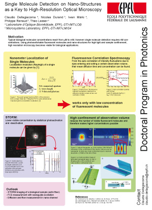
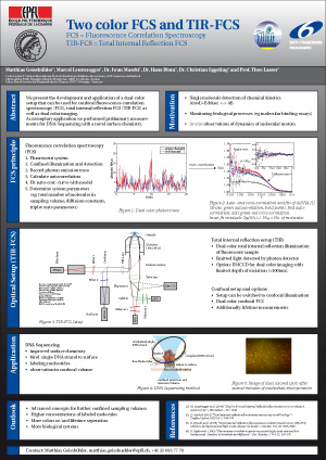
Overview
The detection of single molecules using confocal spectroscopy is of major interest and tremendous improvements have been made within the last decade. The underlying basics however, a pinhole and a single excitation volume element, have remained the same as have the technical limitations.
Our group tries to develop new solutions in order to seek and find new applications in biology, medicine and drug development. The fairly high signal-to-background ratio which is achieved with this technique, paves the way for numerous applications and leaves room for improvement in the realms of the instrumental components ; optical components, illumination or excitation light sources and detectors.
Posters
 |
 |
| – | Details and PDF of poster |
Project: EPFL STI Seed Fund
| Title | Nanostructured surfaces for single molecule detection |
| Funding | EPFL STI Seed Fund 2007 and EPFL STI Seed Fund 2008 |
| Schedule | 01.07.2007 – 30.06.2008 and 01.07.2008 – 30.06.2009 |
| Abstract |
This project combines single molecule analysis with an optical nanostructured platform for enhancing the fluorescence response. The proposed approach provides an important step towards fully integrated, miniature and functional biochips with a particular emphasis on single molecule detection. In addition, new imaging techniques such as structured light illumination for high-resolution microscopy could be integrated and developed. Further, the planar platform opens the possibility for a high degree of parallelization for future high throughput projects. The biomedical optics laboratory is actually involved in a European consortium on Single Molecule Detection for DNA sequencing and phenotyping. This Integrated Project would highly benefit on these preparative research activities. |
Project: FuSyMem
| Title | Functionalized Synthetic Membranes for GPCR Based Sensing |
| Funding | EU Project N° 583143 |
| Schedule | 01.10.2006 – 30.9.2009 |
| Website | http://fusymem.epfl.ch/ |
| Abstract |
We are investigating functionalized synthetic membranes (FuSyMEM) in an international research project funded by the 6th European framework program (FP6). Based on the role they play in events such as sensory perception, drug action and signal recognition, cellular membranes form a versatile area of research. Understanding and controlling cell membrane behaviour relies heavily on having the ‘right tools for the job’. FuSyMEM will provide valuable insight into the membrane protein family, a key class of macromolecules. The project partners aim to utilise the knowledge gained from FuSyMEM to develop innovative applications, such as biosensors, mainly in the pharma-industry. |
Project: Nanoscopic Imaging
| Title | Nanoscopic imaging with optically trapped nanoparticles |
| Funding | FNS Project N° 511153 |
| Schedule | 01.12.2007 – 30.11.2010 |
| Abstract |
Our ability to understand the structures and functions of living systems on a cellular and molecular level is mostly determined by the availability of imaging techniques. Accessing subcellular structures at nanoscopic spatial resolution as well as providing functional information on molecular systems in vivo is the key for the fundamental understanding of cell life. While optical methods provide non-invasiveness, their spatial resolution is limited by a fundamental diffraction limit revealed more than 150 years ago by Ernst Abbe. Overcoming this fundamental limit and accessing in more details cell and membrane structures is the key to access of cell structure and function. This research project addresses this grand-challenge and suggests a novel way of minimally invasive nanoscopic optical imaging inside membranes, vesicles and finally living cells. |