Overview
Optical Coherence Imaging evolved to a new imaging concept with unique features.
Our recent research projects dedicated to Optical Coherence Tomography and in particular to Fourier Domain Optical Coherence Tomography exploit fully these features for fast 3D-imaging with high penetration depths. Several medical applications such as retina imaging, bloodflow monitoring, skin structure and advanced concepts in microscopy with new applications in oncology, diabetes and stem cell research have been successfully addressed.
Posters
 |
 |
| Details and PDF of poster | Details and PDF of poster |
Project: Beta.Image
| Title | Beta.Image – Use of beta-cell imaging in diabetes mellitus |
| Funding | EU Project N° 587067 |
| Schedule | 01.10.2009 – 30.09.2012 |
| Website | http://www.betaimage.eu/ |
| Abstract |
The development of sensitive, non-invasive methods for the characterisation and quantification of beta-cell mass would greatly enhance our means for gaining understanding of the pathophysiology of diabetes and allow the development of novel therapies to prevent, halt and reverse the disease. The aim of this project is to develop and apply innovative approaches for beta-cell imaging, the emphasis being on beta-cell mass regulation (loss and neogenesis) with the perspective of entering initial clinical trials. For this purpose, our approach is to:
To achieve these ambitious goals, we have established a highly interdisciplinary and interactive project. In this way, a unique expertise is achieved regarding tracer development and imaging, beta-cells/diabetes and target definition. |
Project: Functional Imaging
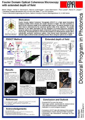
| Title | Functional Imaging and Fourier Domain Optical Coherence Microscopy |
| Funding | FNS Project N° 511358 |
| Schedule | 01.10.2008 – 30.09.2010 |
| Abstract |
The assessment of dynamic processes opens new horizons in biomicroscopy due to the fast 4D-imaging. Monitoring of blood flow, imaging of muscle contractions with the real perspective of monitoring neuronal activity offers a huge untapped potential due to the outstanding performance of xfOCM. In this project we intend to push the limits towards higher resolution and towards phase imaging based on new interferometer concepts. Therefore we will put an additional effort into the development of design tools (focus field engineering, interferometer design including high NA optics) which will together with new concepts in image processing result in an overall improvement of the 3D image quality. |